
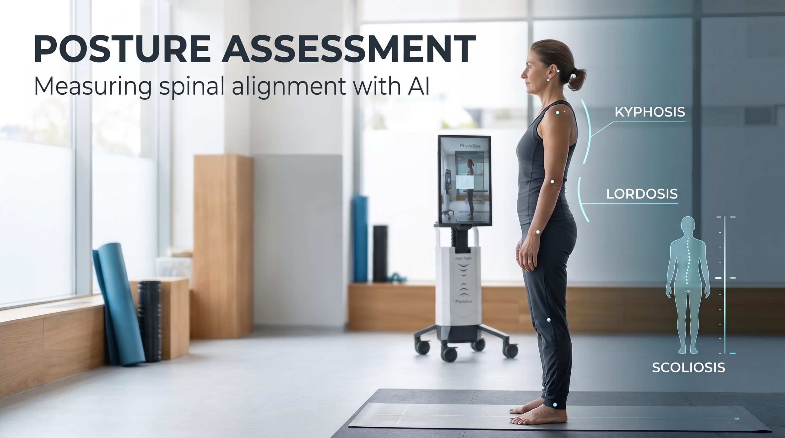
Posture Assessment: How AI Measures Lordosis, Kyphosis, Scoliosis and Spinal Alignment with PhysioEye
In physical medicine and rehabilitation, evaluating the human spine is fundamental to understanding musculoskeletal function, movement efficiency, and physical strain. Clinicians continually evaluate how postural shifts contribute to functional decline, pain syndromes, and movement limitations. However, traditional visual evaluation faces a persistent challenge: human eyes struggle to record sub-millimeter anatomical positions consistently across appointments, clinicians, or different clinical environments.
When a physical therapist or rehabilitation specialist documents a patient’s posture, they evaluate observable physical alignment. It is crucial to recognize that an unusual external appearance alone does not establish a primary spinal disorder or explain the origin of a patient’s pain. To establish meaningful clinical baseline profiles and track recovery, physical medicine requires structured, objective, and repeatable measurement protocols.
According to research indexed by the National Institutes of Health (NIH), standardized physical screening and objective kinematic quantification are essential to eliminate inter-examiner variability and establish reliable clinical documentation.
AI-based posture assessment utilizes optical computer vision and depth sensing to quantify external spinal contours and anatomical landmarks in 3D space. By measuring lordosis, kyphosis, and scoliosis dynamics without physical markers, platforms like PhysioEye deliver repeatable clinical metrics that support longitudinal monitoring and objective care planning.
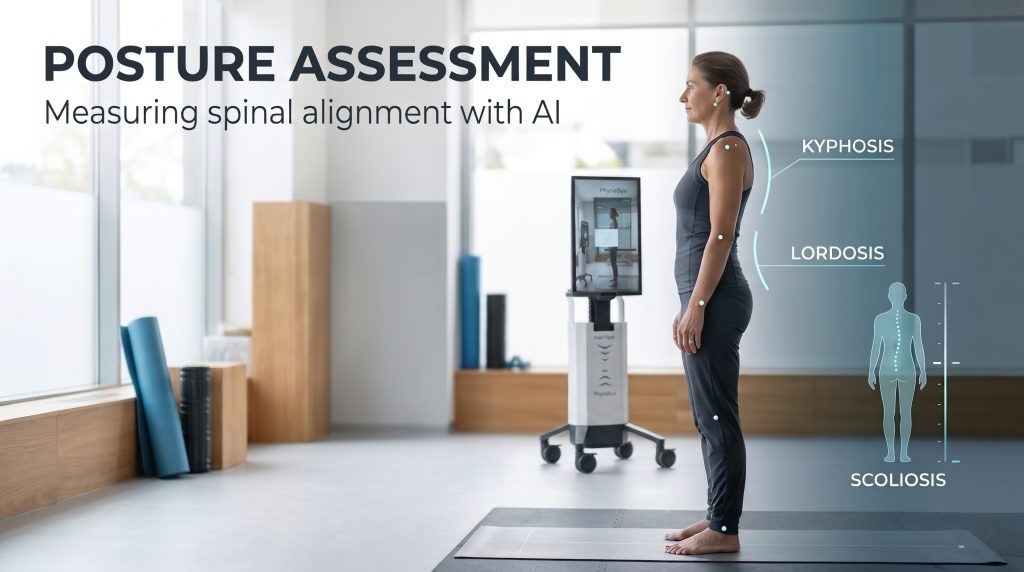
Posture Assessment: What Does Spinal Alignment Tell Us?
Posture assessment describes observable body alignment in static standing and dynamic movement. In standard physical therapy and rehabilitation workflows, capturing subtle changes in posture provides a window into how a patient manages gravitational loads, counterbalances muscular imbalances, and executes fundamental movement patterns.
The primary clinical problem in traditional practice is that visual descriptions—such as “moderate upper-back rounding” or “slight lumbar arching”—are inherently subjective. A description written by one therapist may be interpreted differently by another six weeks later, making objective longitudinal comparison nearly impossible.
Structured posture assessment provides a standardized baseline. However, clinicians must separate anatomical observation from clinical pathology: external postural variations are extremely common across asymptomatic populations, and an observed deviation must always be interpreted alongside clinical symptoms, range of motion, and physical performance testing.
The Three Main Spinal Curvature Patterns
Understanding postural evaluation requires defining the three primary anatomical curvature patterns of the human vertebral column:
-
Lordosis: The natural inward anteroposterior curvature of the cervical and lumbar regions of the spine. When evaluating lumbar lordosis, clinicians look for observable hyper-lordosis (excessive inward arching) or hypo-lordosis (flat-back alignment), both of which alter pelvic tilt and hip mechanics.
-
Kyphosis: The natural outward posterior curvature of the thoracic and sacral regions. While a sagittal thoracic curve is anatomically physiological, excessive thoracic kyphosis (often termed hyper-kyphosis) restricts shoulder girdle mobility, alters respiratory mechanics, and increases cervical strain.
-
Scoliosis: A complex, three-dimensional spinal deformity that involves lateral curvature in the frontal plane, rotational twisting of the vertebrae in the transverse plane, and alteration of sagittal curves. True scoliosis is not simply a sideways tilt seen in a 2D photograph; it involves 3D vertebral rotation and rib cage asymmetry.
It is essential to emphasize that normal spinal curvature varies significantly among healthy individuals based on age, sex, ethnicity, and body composition. Crucially, surface-based external posture measurements assess skin contours and palpable anatomical landmarks—they are not anatomically identical to internal radiographically measured Cobb angles or internal vertebral rotation.
How Clinicians Assess Spinal Posture Today
Modern rehabilitation clinics employ a spectrum of diagnostic approaches to evaluate spinal posture, ranging from simple visual inspection to sophisticated optical imaging:
-
Visual Inspection and Plumb-Line Assessment: The clinician visually inspects the patient against a vertical plumb line or grid chart, checking for shoulder height differences, pelvic tilts, and head protraction.
-
Palpation and Surface Measurements: Utilizing tools like scoliometers, inclinometers, or flexible rulers, clinicians measure skin surface angles over specific spinous processes.
-
Photographic and Photogrammetric Methods: Capturing 2D photos from anterior, posterior, and lateral views allows therapists to manually draw lines and calculate two-dimensional joint angles using software.
Systematic reviews archived on PubMed Central demonstrate that while surface measurements show moderate-to-strong intra-examiner reliability when strict protocols are enforced, inter-examiner agreement varies widely based on examiner experience, anatomical landmark palpation accuracy, and small changes in resident standing posture.
Why Visual Assessment Can Be Difficult to Standardize
Relying purely on unassisted visual assessment introduces multiple variables that hinder long-term clinical standardization:
-
Observer Variability: Different clinicians emphasize different visual cues. What an occupational therapist categorizes as “mild thoracic slump,” an orthopedic surgeon might document as “normal variation.”
-
Inconsistent Positioning: Minor changes in a patient’s stance width, weight distribution, or footwear between appointments drastically alter the visual presentation of the lumbar curve and shoulder alignment.
-
Landmark Identification Shifts: Without fixed, objective spatial references, identifying exact spinous process levels relies on subjective touch, which changes across different clinical evaluators.
Recognizing these limitations does not mean visual observation is useless. Experienced clinical observation remains a vital component of physical examination; however, pairing systematic visual observation with repeatable, objective measurement tools creates a far more trustworthy clinical record.
How AI-Based Spinal Posture Assessment Works
Artificial intelligence and computer vision transform how external body alignment is measured. Rather than relying on manual goniometers or subjective visual estimates, AI systems execute a structured, mathematical workflow:
-
Optical Image and Depth Acquisition: A specialized camera setup captures the patient’s external body stance from defined angles.
-
Computer Vision Landmark Detection: Deep learning models identify key anatomical landmarks (such as acromion processes, anterior superior iliac spines, C7/T1 junctions, and auditory meati) across the body surface.
-
Algorithmic Spatial Derivation: Algorithms calculate specific angular relationships, surface asymmetry indices, and deviation metrics across the sagittal, frontal, and transverse planes.
-
Standardized Longitudinal Recording: Measurements are stored in an objective digital format, allowing future sessions to be overlaid and compared under identical calibrated conditions.
It is critical to distinguish two-dimensional (2D) photographic analysis from three-dimensional (3D) depth-based analysis. As demonstrated in optical research indexed by Springer Nature, 3D body representations incorporate volumetric spatial depth, capturing subtle rotation and postural sway that 2D photos completely miss. However, even a high-resolution 3D surface model measures skin geometry—it does not directly visualize internal bone structure beneath subcutaneous fat and tissue.
Can AI Measure Lordosis, Kyphosis and Scoliosis?
To evaluate AI posture tools objectively, clinicians must understand the exact capabilities and limitations of camera-based algorithms for specific spinal configurations:
Lordosis and Kyphosis
AI algorithms can accurately quantify external surface contours, calculating sagittal angles that reflect lumbar curvature and thoracic rounding. These external measurements correlate well with postural changes during functional movement. However, surface-calculated curves must never be presented as mathematically identical to radiographic vertebral body angles (such as sagittal Cobb angles derived from standing X-rays) unless specific, population-validated transformation models have been applied.
Scoliosis
Computer vision excels at detecting surface asymmetry, such as shoulder height discrepancies, scapular prominence, waist contour asymmetry, and rib humps during forward-bending tasks. While AI provides a sensitive, zero-radiation method to document visible asymmetric features associated with scoliosis, it cannot confirm structural vertebral rotation, establish Cobb angle severity, or replace diagnostic standing radiographs when structural scoliosis is suspected.
Across all three conditions, an AI system’s clinical performance depends entirely on its specific hardware calibration, landmark estimation models, training dataset diversity, and validation against reference gold standards. Evidence validating automated measurement from spinal X-rays cannot be used to justify claims that an optical camera achieves the same internal diagnostic precision.
What Makes an AI Posture Measurement Clinically Useful?
A posture measurement that looks precise on a digital screen is worthless if it lacks clinical validity and reliability. For an AI posture assessment tool to earn clinical trust, it must demonstrate rigorous psychometric properties:
| Metric Parameter | Clinical Meaning | Requirements for Trustworthy Measurement |
| Validity | Does the tool measure what it claims to measure? | Must be validated against validated gold standards (e.g., 3D motion capture) |
| Test-Retest Reliability | Does it produce identical results under unchanged conditions? | High Intraclass Correlation Coefficients (ICC > 0.90) |
| Minimal Detectable Change (MDC) | The smallest real change beyond measurement error | Must differentiate true physical progress from system noise |
| Standardized Positioning | Consistent patient stance and distance | Fixed stance width, calibrated camera distance, clear joint visibility |
Clinicians must look for systems that demonstrate repeatable measurement performance across diverse body types, clothing layers, and standing positions, ensuring that observed changes reflect genuine clinical progress rather than environmental measurement noise.
From a Single Snapshot to Longitudinal Posture Monitoring
The true clinical value of objective posture assessment lies not in a single baseline measurement, but in tracking long-term trends throughout a rehabilitation program.
When a patient undergoes a multi-week physical therapy intervention aimed at improving postural control or managing hyper-kyphosis, repeated objective measurements allow clinicians to evaluate whether prescribed exercise protocols are yielding structural and functional adaptations.
However, clinicians must interpret longitudinal data within context: a measured decrease in thoracic surface rounding does not automatically equal complete symptom resolution, just as a minor structural deviation does not imply clinical deterioration. Objective posture data must always be correlated with functional outcome measures, such as Gait Speed Measurement, the Functional Reach Test, and patient-reported pain scores.
How PhysioEye Fits into AI-Based Posture Assessment
At Hash-Tech GmbH, our biomedical engineering approach focuses on delivering validated, optical kinematic intelligence. The PhysioEye platform delivers Class Im CE-marked 3D Markerless Motion Capture without requiring physical body markers, force plates, or wearable sensors.
Driven by vision models like ViTPose, PhysioEye captures high-density 3D spatial coordinates during an Automated Mobility Assessment. When evaluating posture, PhysioEye documents external spinal alignment patterns, shoulder-pelvis symmetry, and multi-planar trunk orientation.
By removing human error from landmark identification, PhysioEye supports standardized clinical workflows, making longitudinal comparisons accurate and repeatable across long-term therapy programs.
When postural deficits or axial core weakness require physical intervention, clinicians integrate diagnostic insights from PhysioEye with targeted rehabilitation delivered by the ErgoBot system. ErgoBot is engineered specifically as a stationary upper- and lower-limb rehabilitation robot for all joints—it is definitively not a wearable exoskeleton, gait suit, or strength training machine.
Patients remain comfortably seated while connecting to ErgoBot via minimal, clinically accurate ring-shaped attachments. This stationary setup allows for high-dose Robotic Assisted Ergotherapy / Occupational Therapy and Passive Range of Motion Therapy to strengthen core stabilizers, support Joint Stiffness Prevention, and promote Neuromuscular Re-education without exposing frail patients to over-ground fall risks.
PhysioEye is designed to document external kinematic alignment and posture patterns accurately. It does not independently diagnose structural scoliosis or claim to replace internal radiographic X-ray measurements—it serves as an objective, zero-radiation clinical decision support tool for physical medicine professionals.
The Future of Spinal Posture Assessment
Spinal posture evaluation is shifting away from subjective visual guesses toward standardized, documented, and repeatable digital analytics. The goal of integrating artificial intelligence into physical therapy and orthopedics is not to replace human clinical reasoning or medical imaging. Instead, AI serves as an objective clinical partner—supplying therapists with precise, quantitative movement data that enriches physical examinations.
As optical computer vision technology continues to advance, zero-touch 3D motion tracking will become a seamless component of routine physical screening across outpatient clinics, rehabilitation centers, and Nursing Home Automation / Elderly Care Solutions. By combining markerless diagnostic assessments with safe, stationary robotic ergotherapy, healthcare providers can deliver transparent, evidence-based care that optimizes musculoskeletal health across all age groups.
Frequently Asked Questions
What is spinal posture assessment? Spinal posture assessment is the systematic clinical evaluation of the alignment, curves, and spatial orientation of the spine and surrounding anatomical structures (such as the pelvis and shoulders) during static standing and dynamic movement.
What is the difference between lordosis and kyphosis? Lordosis refers to the natural inward curvature of the cervical and lumbar spine (sagittal plane), whereas kyphosis refers to the natural outward curvature of the thoracic and sacral regions. Both are physiological curves, but excessive lordosis (hyper-lordosis) or excessive kyphosis (hyper-kyphosis) can alter biomechanical loading.
How is scoliosis different from ordinary postural asymmetry? Ordinary postural asymmetry often stems from habitual muscle imbalances or leg length discrepancies without vertebral structural changes. Scoliosis is a complex three-dimensional structural spinal deformity involving lateral curvature, axial rotation of the vertebrae, and rib cage asymmetry.
Can AI measure spinal curvature from a camera image? AI can measure external surface curvature, skin contours, and landmark locations from camera images. However, camera-based AI measures surface geometry, which reflects underlying spinal shape but is not identical to internal radiographically measured vertebral angles.
Can camera-based posture assessment replace spinal X-rays? No. Camera-based posture assessment evaluates external surface alignment and skin geometry without radiation. It cannot visualize internal bone structure, confirm structural vertebral rotation, or calculate Cobb angles, meaning diagnostic X-rays remain necessary when structural spinal pathology is suspected.
How accurate is AI-based spinal posture assessment? Accuracy depends on the specific AI algorithm, camera technology (2D vs. 3D depth), patient positioning protocol, and validation evidence. High-quality 3D markerless systems achieve excellent test-retest reliability for surface parameters when standardized clinical protocols are followed.
Can AI posture assessment track changes over time? Yes. One of the greatest strengths of AI posture assessment platforms like PhysioEye is longitudinal tracking. By maintaining consistent camera distances and patient positioning, the system allows clinicians to compare baseline surface measurements against post-intervention data to monitor alignment trends objectively.