
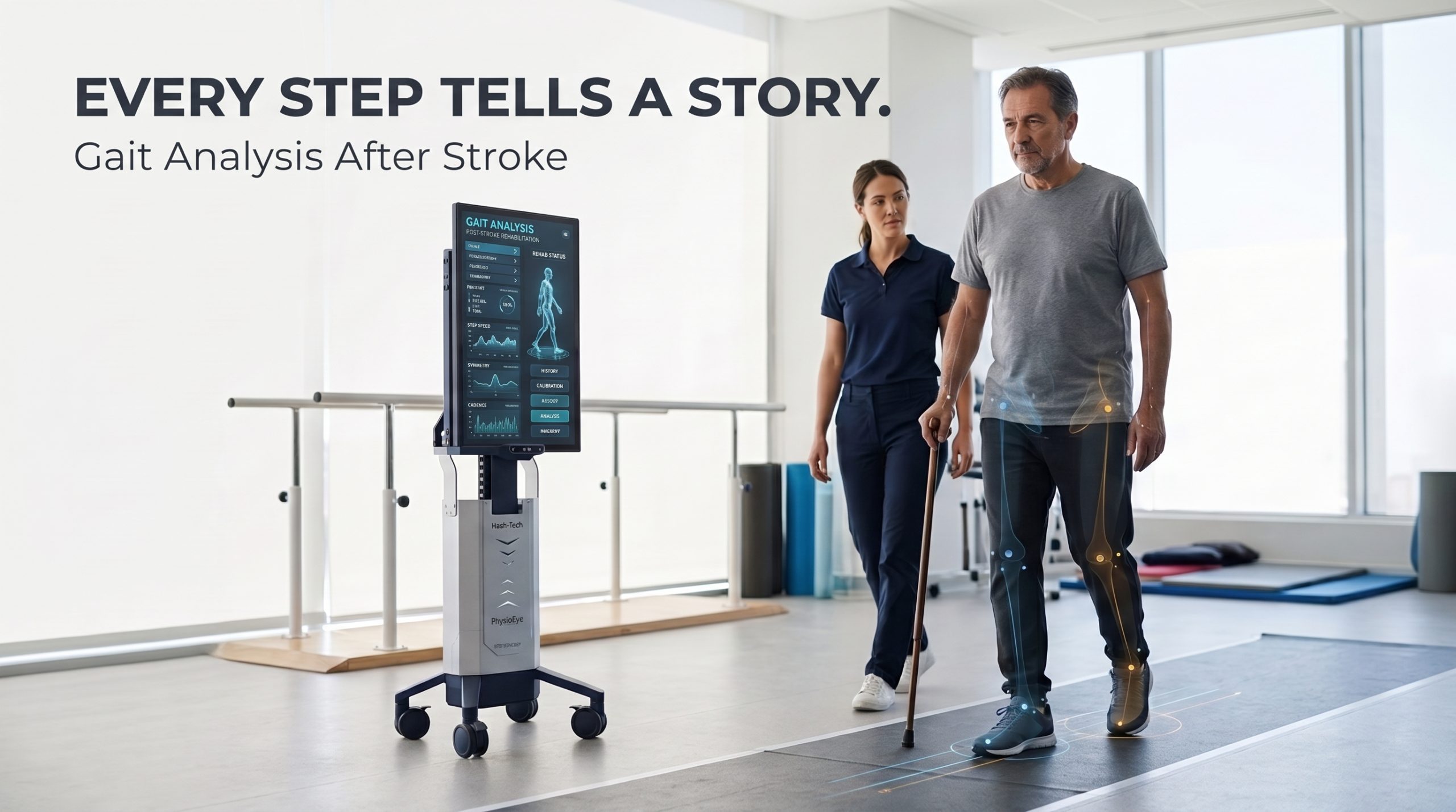
Gait Analysis After Stroke: 8 Key Metrics to Track Walking Recovery
Restoring independent ambulation is one of the highest-priority goals for stroke survivors and their rehabilitation teams. Following a cerebrovascular accident, neurological disruptions in motor control, sensory integration, and muscle activation often manifest as significant gait impairments. However, gait recovery is rarely linear, and two stroke survivors with similar walking speeds may exhibit vastly different underlying biomechanical strategies and movement efficiencies.
To construct evidence-based rehabilitation plans and track recovery objectively, physical medicine relies on structured gait analysis after stroke. Rather than relying solely on visual observation or single functional benchmarks, quantitative gait characterization measures both overall walking capacity and specific movement quality parameters.
According to global clinical practice consensus published in PubMed Central, integrating standardized outcome measures and biomechanical assessments is essential to differentiate true neural recovery from compensatory motor strategies.
Gait analysis after stroke quantifies walking recovery across eight core biomechanical metrics: walking speed, endurance, step length, cadence, gait-cycle timing, spatial/temporal asymmetry, paretic propulsion, and lower-limb joint kinematics. Utilizing a multi-metric diagnostic framework paired with markerless 3D motion analytics like PhysioEye allows clinicians to isolate specific motor deficits, measure true movement quality, and tailor personalized rehabilitation pathways.
What Changes After Stroke?
A stroke frequently damages corticospinal pathways and motor cortex circuitry, resulting in post-stroke hemiparesis—weakness or altered voluntary motor control on one side of the body. These neurological deficits create substantial variation in walking patterns across individuals:
-
Paretic vs. Non-Paretic Discrepancies: Reduced voluntary muscle recruitment on the paretic side forces the non-paretic limb to assume a greater workload for weight acceptance and propulsion.
-
Altered Phase Dynamics: The stance phase on the paretic limb is frequently shortened due to instability or pain, while the swing phase is prolonged, causing marked temporal asymmetry.
-
Reduced Ankle Push-Off and Propulsion: Impaired plantarflexor recruitment limits forward propulsive impulse, resulting in reduced walking speed and shorter step lengths.
-
Compensatory Movement Strategies: Patients often adopt compensatory mechanics—such as hip circumduction, pelvic hiking, or excessive trunk leaning—to clear the swing foot and advance the leg.
-
Heterogeneity Across Recovery Stages: Gait characteristics vary significantly between early acute inpatient rehabilitation and chronic stroke states, as well as between individuals with similar functional walking categories.
Crucially, functional walking ability, movement quality, and physical endurance are related but distinct clinical outcomes. A patient may achieve rapid functional ambulation by relying heavily on energetic compensations, yet possess severe biomechanical asymmetry that increases fall risk and long-term joint wear.
8 Key Metrics to Track, Gait Analysis After Stroke
A comprehensive post-stroke gait evaluation examines spatiotemporal, kinetic, and kinematic variables. Below are the eight foundational metrics prioritized by stroke rehabilitation consensus panels.
Walking Speed
Comfortable and maximum gait speed—measured in meters per second (m/s) is widely recognized as the single most practical indicator of overall walking performance and community ambulation status.
-
Clinical Relevance: Gait speed correlates strongly with lower-limb strength, balance confidence, and functional independence. A gait speed under 0.4 m/s typically indicates household ambulation, 0.4 to 0.8 m/s represents limited community ambulation, and >0.8 m/s reflects full community mobility.
-
Standardized Assessment: Evaluated using the 10-Metre Walk Test (10MWT).
-
Limitation: Walking speed alone is a summary metric—it cannot reveal how the speed was achieved or differentiate between true motor recovery and energy-costly compensations.
Walking Endurance
Walking endurance reflects the distance covered during a sustained walking task, integrating cardiovascular fitness, muscular stamina, and fatigue resistance.
-
Clinical Relevance: Short-burst velocity measured during a 10-meter sprint often fails to reflect a patient’s capacity to navigate real-world environments like shopping centers or parks. Endurance assessments expose fatigue-induced gait breakdown.
-
Standardized Assessment: Measured via the 6-Minute Walk Test (6MWT).
-
Influencing Factors: Results depend heavily on fatigue levels, rest breaks, footwear, orthotic use, and standardized hallway protocol enforcement.
Step Length and Stride Length
Step length measures the distance from the point of initial contact of one foot to the initial contact of the opposite foot. Stride length represents a complete gait cycle (initial contact of one foot to initial contact of the same foot).
-
Clinical Relevance: Stroke survivors typically exhibit significantly shortened step lengths on the paretic side, non-paretic side, or both. Differences between sides indicate spatial step asymmetry.
-
Measurement Consideration: Because walking speed directly influences spatial step distance, clinicians must ensure identical walking speed conditions when comparing longitudinal step length measurements.
Cadence
Cadence measures the number of steps taken per minute (steps/min).
-
Clinical Relevance: Cadence interacts dynamically with step length to determine overall walking speed (Speed = Step Length * Cadence). Following a stroke, patients often increase cadence to compensate for reduced step length.
-
Diagnostic Caution: An increase in cadence should never be interpreted as a standalone indicator of recovery. If step length remains severely restricted, a high cadence merely reflects rapid, unstable shuffling.
Gait-Cycle Timing (Phase Dynamics)
The gait cycle consists of stance phase (weight-bearing) and swing phase (unloaded advancement), further broken down into single-limb support and double-limb support durations.
-
Clinical Relevance: Stroke patients frequently exhibit prolonged double-support time (seeking stability) and reduced single-limb support time on the paretic leg due to weight-bearing insecurity.
-
Diagnostic Value: Detailed timing measures uncover subtle neural recovery improvements—such as prolonged single-leg stance on the paretic side—even before overall walking speed changes.
Gait Asymmetry
Asymmetry evaluates side-to-side differences across spatial (step length) and temporal (stance, swing, and double-support times) variables, often quantified using mathematical asymmetry indices.
-
Clinical Relevance: Systematic reviews archived on PubMed Central establish that gait asymmetry reflects underlying motor control deficits and compensatory gait patterns.
-
Biomechanical Nuance: Perfect symmetry is not automatically equivalent to efficient or normal walking; forcing absolute symmetry prematurely in a severely hemiparetic patient can sometimes reduce functional stability.
Paretic-Limb Propulsion
Paretic-limb propulsion measures the affected leg’s active mechanical contribution to forward body acceleration, driven primarily by ankle plantarflexor push-off and hip extension.
-
Clinical Relevance: The Stroke Recovery and Rehabilitation Roundtable consensus explicitly prioritizes kinetic measures of propulsion—specifically stride-normalized ground-reaction forces—to evaluate paretic leg contribution.
-
Diagnostic Distinction: A patient can increase walking speed by over-utilizing the non-paretic leg to pull the body forward. Measuring propulsive impulse separates true paretic recovery from non-paretic compensation.
Lower-Limb Joint Kinematics and Overall Gait Deviation
Joint kinematics map the angular trajectories of the hip, knee, and ankle joints across sagittal, frontal, and transverse planes throughout the gait cycle.
-
Clinical Relevance: Common post-stroke kinematic deviations include stiff-knee gait (reduced peak knee flexion during swing), excessive ankle plantarflexion (foot drop), and reduced hip extension.
-
Summary Scores: When full 3D motion data is available, multivariate indices such as the Gait Profile Score (GPS) or Gait Deviation Index (GDI) summarize total gait deviation from neurotypical control baselines into a single numerical score.
Standardized Clinical Outcome Measures
Beyond instrumented biomechanical metrics, stroke rehabilitation consensus guidelines recommend a core set of standardized outcome tools to establish functional baselines and document progress:
-
10-Metre Walk Test (10MWT): Assesses comfortable and fast walking speed over a short distance.
-
6-Minute Walk Test (6MWT): Evaluates sub-maximal walking endurance and cardiovascular response.
-
Functional Ambulation Category (FAC): Categorizes the level of physical assistance and supervision required for ambulation (ranging from non-functional to independent community walker).
-
Dynamic Gait Index (DGI): Assesses dynamic balance and the ability to modify gait in response to changing environmental demands (e.g., speed changes, head turns).
-
Functional Gait Assessment (FGA): An expanded gait assessment incorporating narrow base-of-support tasks and eyes-closed walking.
-
Timed Up and Go (TUG): Measures functional mobility involving standing up, walking 3 meters, turning, and sitting down.
-
Fugl-Meyer Assessment — Lower Extremity (FMA-LE): Evaluates voluntary lower-limb motor impairment and joint isolation (not a direct walking test).
-
Berg Balance Scale & Mini-BESTest: Complementary balance scales that quantify static and dynamic postural control.
Note: Stroke consensus guidelines highlight that performance-based walking tests like the 10MWT or 6MWT are non-testable in patients requiring physical assistance (FAC < 3), underscoring the need for adaptable assessment tools across recovery stages.
Traditional vs. Technology-Driven Gait Assessment
Post-stroke gait evaluation spans a wide technological continuum, each offering distinct clinical trade-offs:
-
Visual Clinical Observation: Highly portable and zero-cost, but subject to inter-rater variability and incapable of quantifying high-speed spatial-temporal micro-changes.
-
Instrumented Walkways & Pressure Mats: Excellent for capturing spatial-temporal parameters (step length, contact time), but cannot directly track upper-body kinematics or multi-planar joint angles.
-
Wearable Inertial Sensors (IMUs): Provide portable kinematic tracking in real-world environments. However, systematic reviews published in ScienceDirect highlight that wearable measurement properties vary significantly by device and show weaker agreement for swing-time parameters in slow-walking populations.
-
Optical Markerless 3D Motion Capture: Uses computer vision depth sensors to track 3D kinematics without forcing stroke survivors to wear uncomfortable physical markers, sensors, or suit harnesses.
AI and Digital Gait Analysis: Capabilities and Clinical Accuracy
Artificial intelligence and computer vision pose estimation models have revolutionized physical medicine. By processing high-resolution depth and optical data, AI platforms calculate spatiotemporal parameters, track left-right symmetry, and generate automated rehabilitation reports.
Validity, Reliability, and Technical Limitations
For AI gait analysis to earn clinical trust in stroke rehabilitation, algorithms must be specifically validated in post-stroke populations rather than healthy cohorts:
-
Reference System Validation: AI camera systems must demonstrate high concurrent validity against laboratory gold standards (such as optoelectronic marker systems).
-
Sensitivity to Pathological Gait: Algorithms must remain reliable during slow walking speeds (<0.4 m/s), severe asymmetric shuffling, and when patients use assistive walking frames, canes, or ankle-foot orthoses (AFOs).
-
Spatial vs. Temporal Accuracy: Recent 2026 systematic reviews report that markerless camera systems generally demonstrate stronger agreement for spatial parameters (step length, stride width) than for subtle temporal parameters like swing-phase duration in severe hemiparesis.
-
Environmental Standardization: Camera angles, lighting, clothing fit, and walking surface consistency must be controlled to prevent measurement artifacts.
Crucially, digital measurements quantify movement patterns—they do not independently diagnose the underlying neurological root cause. Experienced clinical interpretation remains essential.
How PhysioEye Supports Markerless 3D Gait Assessment
At Hash-Tech GmbH, our biomedical technology vision centers on bringing objective kinematic analysis into everyday clinical workflows. The PhysioEye platform delivers Class Im CE-marked 3D Markerless Motion Capture without attaching physical markers or wearable sensors to the stroke survivor’s body.
Driven by computer vision models like ViTPose, PhysioEye conducts an Automated Mobility Assessment during functional walking tasks. The system automatically measures joint excursion, spatial step symmetry, and gait-cycle phase dynamics, presenting an objective digital baseline that supports repeatable longitudinal tracking.
When kinematic assessment reveals severe lower-limb weakness, spasticity, or joint stiffness, physical therapists integrate diagnostic insights from PhysioEye with targeted motor therapy using the ErgoBot system.
ErgoBot is engineered specifically as a stationary upper- and lower-limb rehabilitation robot for all joints—it is definitively not a wearable exoskeleton, gait suit, or strength training machine. Patients remain safely seated in a standard clinical chair or wheelchair while connecting to ErgoBot via minimal, clinically accurate ring-shaped attachments.
This stationary configuration allows stroke survivors to undergo high-repetition Robotic Stroke Rehabilitation and Robotic Assisted Ergotherapy / Occupational Therapy, supporting Neuromuscular Re-education and lower-limb motor recovery without exposing vulnerable patients to over-ground fall risks.
Complete ErgoBot PhysioEye integration enables clinicians to track how targeted robotic joint mobilization translates directly into improved functional gait metrics over time.
Key Takeaways
-
Multi-Metric Evaluation: Comprehensive post-stroke gait analysis must evaluate functional capacity (speed, endurance) alongside movement quality (asymmetry, propulsion, kinematics).
-
Dissecting Compensation vs. Recovery: Faster walking does not automatically signify complete motor recovery; patients often compensate using non-paretic propulsion or hip circumduction.
-
Standardized Consensus Tools: Clinical pathways should pair instrumented metrics with core consensus tools like the 10MWT, 6MWT, FAC, and DGI.
-
Markerless Diagnostic Safety: Optical 3D platforms like PhysioEye deliver validated kinematic tracking without physical markers or wearable suit hassles.
-
Targeted Stationary Interventions: Interventions using ErgoBot deliver high-dose joint mobilization via minimal ring-shaped end-effectors, supporting neuromuscular re-education with zero fall risk.
Frequently Asked Questions
What is gait analysis after stroke? Gait analysis after stroke is the systematic evaluation of walking mechanics in stroke survivors. It measures spatiotemporal variables (speed, step length, cadence), kinetic forces (paretic propulsion), and joint kinematics (range of motion) to quantify recovery and guide physical rehabilitation.
Which gait parameters are most important after a stroke? While walking speed (10MWT) and endurance (6MWT) are critical functional benchmarks, stroke consensus guidelines emphasize measuring gait asymmetry, paretic limb propulsion, and single-limb stance time to assess true movement quality and neural recovery.
How is walking speed measured after stroke? Walking speed is typically measured using the 10-Metre Walk Test (10MWT), where the time taken to walk a central 6-meter distance (with acceleration and deceleration zones) is recorded to calculate velocity in meters per second ($\text{m/s}$).
What is the difference between the 10MWT and the 6MWT? The 10MWT measures short-burst comfortable and maximum walking speed over a short distance, whereas the 6MWT measures physical endurance and cardiovascular stamina over a sustained 6-minute walking duration.
Why is gait asymmetry common after stroke? Gait asymmetry stems from post-stroke hemiparesis, where impaired voluntary muscle control on the paretic side leads to shortened paretic stance times, altered step lengths, and compensatory reliance on the healthy, non-paretic limb.
Does faster walking mean gait recovery is complete? No. A stroke survivor may walk faster by relying heavily on energetic compensations, such as pulling the body forward with the non-paretic limb or hiking the pelvis, without restoring normal muscle coordination or paretic leg propulsion.
How can gait analysis track rehabilitation progress? By establishing a standardized 3D kinematic baseline at admission, clinicians can re-evaluate the patient at regular intervals under identical walking conditions to objectively track changes in step symmetry, joint excursion, and propulsion.
Can AI measure gait after stroke? Yes. AI-powered computer vision platforms process optical and depth data to measure spatiotemporal parameters, joint angles, and step symmetry without requiring physical markers or attached sensors.
How accurate is markerless gait analysis? High-quality 3D markerless camera systems demonstrate strong validity and reliability when compared against optoelectronic reference standards. Accuracy is strongest for spatial parameters (step length) and requires validation in post-stroke populations.
Can gait analysis identify why a person walks abnormally after stroke? Gait analysis quantifies mechanical deviations (such as reduced knee flexion or stance asymmetry). However, determining the root neurological cause (such as spasticity vs. weakness) requires combining motion metrics with physical clinical examination.